Fibroid Treatment
Fibroid Treatment

If there are no symptoms, treatment is usually not required. In women with significant symptoms, treatment may be medical or surgical.
Medical Treatment
Medications called gonadotropin-releasing hormone (GnRH) analogs (Lupron) are commonly used in the medical treatment of fibroids. They are usually only given as a temporary measure, such as during the time a woman is preparing for surgery to remove the fibroids. GnRH analogs cause a reduction in estrogen levels.
Most women taking these medicines have a cessation of the menstrual period. The lack of periods can help women with anemia from fibroid related heavy or prolonged menstrual bleeding to build their blood counts up before surgery.
In some cases, GnRH analogs can cause shrinkage of fibroids which may allow them to be removed through a smaller incision. The fibroids rapidly enlarge again after the medication is discontinued. Since there are adverse effects from prolonged low estrogen levels, GnRH analogs are only a temporary measure.
Surgical Treatment
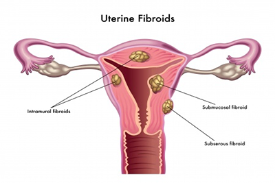
The type of surgery needed is dependent upon the size, number and location of fibroids. In addition, the underlying problem is important.
Obviously, a woman with infertility who wants to keep her uterus would be treated differently than a peri-menopausal woman who is done with her childbearing. Procedures that are performed for women to maintain the capability for childbearing or to improve fertility include:
- Abdominal myomectomy
- Laparoscopic myomectomy
- Hysteroscopic myomectomy
- Uterine artery embolization
Abdominal Myomectomy: Myomectomy means removal of a fibroid. In an abdominal myomectomy, an incision is made through the abdomen to expose the uterus, and the fibroids are excised from the uterine muscle. This approach is most appropriate in women who want to maintain childbearing, and who have multiple fibroids or very large fibroids.
Laparoscopic Myomectomy: In this procedure, the fibroids are removed through laparoscopy . With laparoscopy, a fiberoptic telescope inserted through a tiny incision in or below the belly button, through which the surgeon can visualize the uterus.
Additional tiny incisions are used to introduce long thin operating instruments which can be manipulated to remove the fibroids and repair the uterus afterward.
Laparoscopy is most appropriate for women with one or two small to moderate sized fibroids that are located on the outer surface of the uterus.
Hysteroscopic Myomectomy: As in the case of diagnosing fibroids, hysteroscopy involves placing a fiber optic telescope through the vagina and cervix and into the uterine cavity.
Long thin surgical instruments can be introduced into the uterus using an operative channel in the hysteroscope. Saline is used to keep the walls of the uterus separated.
This procedure can only be done on fibroids that mostly located within the uterine cavity.
Uterine Artery Embolization: Uterine Artery Embolization (UAE) is performed by a radiologist. Using x-ray, a catheter is threaded through blood vessels toward the uterus. Tiny spheres are injected into the blood vessels that feed a fibroid causing the blood supply to be blocked. Without a blood supply, the fibroid will begin to breakdown.
This technique is relatively new. There is very little data on the potential risks it may cause for a woman who subsequently becomes pregnant.
Recently, a multicenter Dutch study looked at the effect of uterine artery embolization on ovarian function. Doctors in the study measured a hormone (anti-mullerian hormone or AMH) which is correlated with ovarian reserve. Women who had UAE had a much more rapid decline in AMH than would have been expected.
This suggests that uterine artery embolization may cause damage to the ovaries and deplete the number of eggs remaining in the ovaries.


